
What tumour-neuron synapses mean for glioblastoma - and why a local siRNA gel is the proposed answer
A 25 August Nature Biomedical Engineering paper shows malignant synapses concentrated at the GBM-brain interface, then tests an intracavitary TrkB-silencing hydrogel that more than doubled median survival in a mouse resection model.
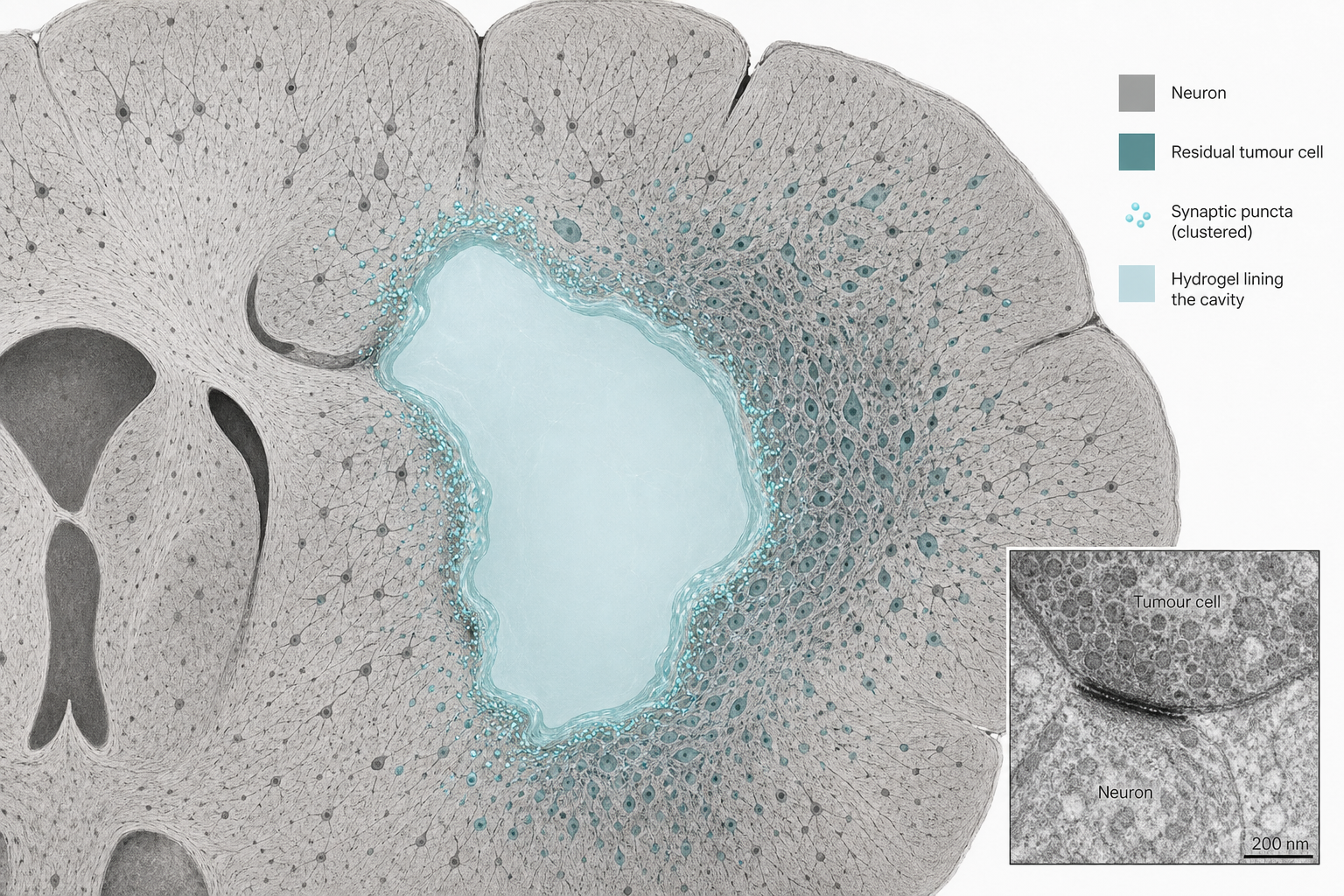
Cancer neuroscience has a delivery problem. Glioma cells form excitatory synapses with neurons, ride BDNF-TrkB and AMPA signalling, and use that activity to invade, to resist radiotherapy, and to remodel the circuits that cognition depends on. The same synapses keep cortex alive. Systemic AMPA antagonists and pan-Trk inhibitors therefore collide with the organ they are trying to spare. A paper published on 25 August 2026 in Nature Biomedical Engineering treats that collision as a spatial problem rather than a medicinal-chemistry one. Malignant synapses, the authors show, are concentrated at the tumour-brain interface , the rim that surgery leaves behind, and the site of more than 85 percent of recurrences. Their answer is not another brain-penetrant kinase inhibitor. It is an siRNA against NTRK2 (TrkB), packed in a glutathione-responsive nanocarrier, sitting in a pH-sensitive hydrogel in the resection cavity.
That gel is not clinically available. There is no human therapeutic trial, and no FDA discussion in the paper. This work is preclinical. “Clinically translatable” is the authors’ language for a mouse-and-xenograft result. The result is still the cleanest recent attempt to turn a review-article insight , tumour-neuron synapses , into a device that can be put where the recurrences start.
The interface, not the core
The human evidence is anatomical and small. The authors analysed en-bloc glioblastoma specimens from six patients, staining Nestin, NeuN, the presynaptic marker Synapsin-1 and the postsynaptic marker PSD95 across necrotic core, tumour-brain interface, distant cortex and distant white matter. Nestin-positive tumour cells were enriched at the interface. Neuronal somata were sparse there. Synapsin-1 and PSD95 were not. Co-localized puncta , putative excitatory synapses , were significantly more numerous at the interface than in the necrotic core. Public single-cell RNA-seq of peritumoural brain versus tumour core showed the same geography in transcription: synapse-associated genes, calcium channels and glutamate receptors up at the edge, and NTRK2 with them.
TrkB is the hinge. BDNF-TrkB signalling promotes synapse assembly and activity-dependent strengthening through CaMKII, AMPA-receptor trafficking and calcium influx. Systemic Trk inhibitors, including the NTRK-fusion drugs larotrectinib and entrectinib, have limited utility in wild-type GBM precisely because Trk receptors are expressed throughout the central nervous system; ataxia and sensory disturbance are not off-target accidents, they are on-target effects in the wrong cells. This paper is not a fusion-tumour study. It is an attempt to silence wild-type TrkB on residual tumour cells at the cavity rim, and to leave contralateral cortex alone.
A gel that waits for acid and glutathione
The construct is HPAC-siNTRK2@Gel: a hyperbranched poly(amido amine) nanocarrier, disulfide-linked so it fragments in tumour-cell glutathione, PEGylated and capped with cRGD to bind integrin αvβ3 on GBM cells, complexed with siRNA against NTRK2, then locked into an oxidized hyaluronic acid / carboxymethyl chitosan hydrogel. The gel injects through a narrow needle and degrades as extracellular pH falls from 7.4 toward the acidity of recurrent tumour. The logic is sequential: the gel stays put in a CSF-washed cavity; acidity from recurrence releases the nanoparticles; cRGD selects tumour over astrocyte; glutathione releases the siRNA; TrkB falls on the cells that would otherwise re-form the interface synapses.
In the syngeneic GL261 resection model, that sequence more than doubled median survival: 67 days versus 32 days, with more than 30 percent of treated animals alive beyond 90 days. Controls , PBS, free nanocomplex without gel, gel alone, negative-control siRNA gel, non-targeted carrier , did not. MRI showed less cavity-adjacent recurrence; TUNEL and Ki67 showed more apoptosis and less proliferation at day 15. Patient-derived GSC xenografts spanning mesenchymal (G0 147), proneural (GSC2), classical (G0 927) and mixed (GSC1203) subtypes also lived longer after the gel than after surgery alone. Combination with radiotherapy reduced invasive growth at the interface; radiation alone, in the G0 147 model, increased detached tumour-cell clusters, an effect the gel abrogated.
The head-to-head with systemic entrectinib is the comparison the delivery argument needs. In the same GL261 resection setting, local gel and oral entrectinib (120 mg/kg daily for 15 days) suppressed early tumour burden to a similar degree. Survival diverged: median 53 days with the gel versus 28.5 days with entrectinib. The gel knocked down TrkB next to the cavity and spared the contralateral cortex; entrectinib reduced contralateral TrkB and produced motor-coordination deficits and transient liver-enzyme elevations the gel did not. Same target, different address, different toxicity.
What the electrophysiology does not universalize
Patch-seq in GFP-labelled GL261 cells found fewer and smaller spontaneous excitatory postsynaptic currents after gel treatment, with downregulation of Ntrk2 and a synapse-associated gene score that no longer tracked EPSC amplitude. The effect is not uniform across human subtypes. Mesenchymal G0 147 xenografts lost sEPSC frequency but gained amplitude. Proneural GSC2 lost both. Mixed GSC1203 lost frequency only. Classical G0 927, the authors report, showed no significant change in sEPSC frequency or amplitude. Survival still improved in that model. Synaptic dependency is therefore not a universal GBM property, and the paper does not claim it is. A therapy aimed at tumour-neuron synapses will, on this evidence, hit some residual cells harder than others.
The cognitive data should be read with the same restraint. A 16-patient, single-centre cohort of high-grade frontal gliomas was used for lesion-symptom mapping: peripheral (interface) lesions associated more with MoCA and HAMD impairment than core lesions. That is associative, n=16. In GL261 mice, the gel reduced anxiety-like behaviour and improved memory assays without impairing locomotion. Those are mouse tests. They do not make the gel a cognitive therapy.
The translational limit
GL261 is a widely used and imperfect glioblastoma model. Patient-derived GSCs in NSG mice are closer to human tumour biology and further from a human immune microenvironment. Survival doublings in mice do not translate one-to-one. Ethics approvals , Sun Yat-sen University Cancer Center B2024-840-01 for the specimens and cognitive cohort, L102042024040B for the animals , locate the work; they do not license a clinical protocol. Gliadel wafers remain the only FDA-approved intracavitary GBM treatment; they release carmustine, not an siRNA, and they are not synapse-targeted.
What the paper does change is the frame. If the therapeutically relevant synapses sit at a rim that surgery already opens, then the failure of systemic synaptic inhibitors is not only a toxicity problem. It is a geometry problem. An siRNA gel that waits for the acidity of recurrence is one proposed geometry. It will have to survive manufacturing, a first-in-human cavity study, and the subtype heterogeneity the authors themselves documented. Until then it is a mechanism, a mouse survival curve, and a warning not to treat TrkB at the GBM interface as if it were an NTRK fusion in a lung mass.